Scroll this catalogue and the receptor names pile up: GLP-1 receptor, ghrelin receptor, GHRH receptor, melanocortin receptors, calcitonin receptor with RAMPs. They sound like a list of unrelated targets. They are not — almost all of them are G protein-coupled receptors, and several sit in the same subfamily. Understanding the shared architecture explains why peptides are the ligands for these targets, why selectivity is so hard to engineer, and why "agonist" is a less complete description than it sounds.
What is a GPCR?
A protein that threads through the cell membrane seven times, leaving a portion outside the cell and a portion inside. Something binds on the outside; the receptor changes shape; that shape change activates a G protein on the inside; the G protein sets off a cascade.
They are the largest receptor family in the human genome — several hundred of them — and they are the target of a very large share of all drugs. Light, odours, adrenaline and most peptide hormones all signal through one.
The families are conventionally split into classes. Two matter here:
- Class A — the largest, mostly small ligands binding inside the transmembrane bundle. The ghrelin receptor (which ipamorelin targets) and the melanocortin receptors are class A.
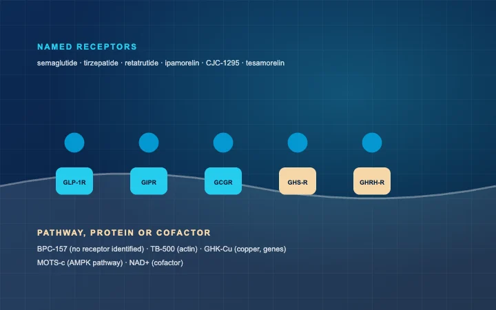
- Class B — the peptide hormone receptors. GLP-1, GIP, glucagon, GHRH and calcitonin receptors are all class B, which means most of the metabolic and growth-axis catalogue points here.
Why does class B suit peptides?
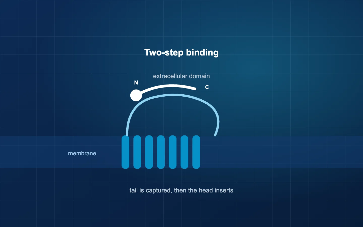
Because of a structural feature the class A receptors lack: a large extracellular domain sitting above the membrane.
Parthier and Stubbs describe the resulting two-step binding model. The peptide's C-terminal region is captured first by that extracellular domain — a relatively long stretch of surface making contact along its length. That tethering brings the peptide's N-terminal region into position to insert into the transmembrane core, and that is what triggers the conformational change.
Two consequences follow, and both show up in this catalogue.
Fragments work. If the extracellular domain recognises one region and the core responds to another, a fragment containing the right parts can be active. GHRH is 44 residues; sermorelin is the first 29 and retains full activity, because residues 30-44 were not doing binding work. Our sequence post covers how these fragments are written.
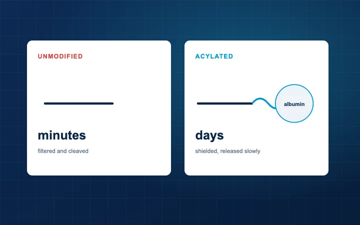
Modifications away from the business end are tolerated. Acylation attaches a fatty chain for half-life, and it works because the attachment point is chosen to sit away from the receptor-facing surfaces. That is the design covered in our acylation post.
| Class A | Class B | |
|---|---|---|
| Typical ligand | Small molecules, short peptides | Peptide hormones, 27-44 residues |
| Extracellular domain | Small | Large, does initial capture |
| Binding | Mostly within the transmembrane bundle | Two-step: tether, then insert |
| In this catalogue | Ghrelin receptor, melanocortin receptors | GLP-1, GIP, glucagon, GHRH, calcitonin |
| Small-molecule agonists | Common | Difficult |
Why are peptide agonists hard to replace?
This is the practical upshot of all that surface area. A class B receptor's binding site is spread across an extracellular domain and a transmembrane core, and a peptide's job is to satisfy both. A small molecule has a fraction of the contact area available to it, so reproducing the full agonist effect is genuinely hard.
That is a large part of why the metabolic field runs on injected peptides rather than tablets — and why Lundquist and colleagues' work on peptide absorption across the intestine is a live research area rather than a solved problem.
What does "agonist" leave out?
More than most product pages suggest. A single receptor can couple to more than one G protein — Gs raising cyclic AMP, Gq mobilising calcium, Gi lowering cAMP — and can additionally recruit β-arrestin, which both dampens signalling and starts its own.
So "compound X is an agonist at receptor Y" is incomplete without saying which pathway was measured. Two compounds can both be agonists and produce different downstream balances, which is what the literature means by biased agonism.
For a study design, three consequences:
- Your readout defines your result. A cAMP assay and a β-arrestin recruitment assay can rank the same two compounds differently, and neither is wrong.
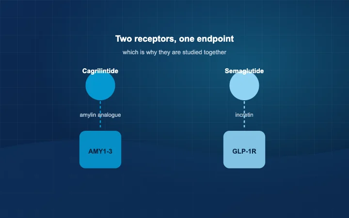
- The expression system matters. Which G proteins a cell line carries shapes what you can detect. The amylin case is the sharpest version — the calcitonin receptor gives amylin pharmacology only when the right RAMP accessory protein is present, which our amylin post covers.
- Selectivity claims need the panel. Saying a compound is selective means it was tested against the alternatives, as our melanocortin post discusses for the melanotan family.
Frequently asked questions
Do all the compounds here act on GPCRs?
No. BPC-157 has no established single receptor; TB-500 acts on actin, a structural protein; NAD+ is a cofactor; SS-31 binds cardiolipin, a membrane lipid. The receptor-and-target table in our reference post maps which is which.
Is the ghrelin receptor really the same family as a light receptor?
Structurally, yes — rhodopsin is the archetypal class A GPCR and the ghrelin receptor is class A too. Seven transmembrane helices and a G protein underneath, with wildly different things binding on top.
What are RAMPs?
Receptor activity-modifying proteins. They partner with a receptor and change what it recognises. The calcitonin receptor plus RAMP1, 2 or 3 produces the AMY1-3 amylin receptor phenotypes — same gene, different pharmacology.
Why does this matter if I am only doing binding assays?
Because binding and activation are different measurements. A compound can bind well and activate poorly, or bind one site while the functional response comes from another. Reporting affinity as if it were potency is a common slip, and our methods post asks for the method alongside every figure.
References
- Parthier C, Stubbs MT. Peptide hormone recognition in class B GPCRs: role of the extracellular domain. In: Drug Discovery. Royal Society of Chemistry, 2011:75-99. doi.org/10.1039/9781849733441-00075
- PubChem, National Center for Biotechnology Information — compound records for the ligands referenced. pubchem.ncbi.nlm.nih.gov
- Bardhan M, et al. Polymorphism of melanocortin receptor genes — association with inflammatory traits and diseases. Diseases 2025;13(9):305. doi.org/10.3390/diseases13090305
Every product mentioned is sold for laboratory research use only and is not for human or animal use. Nothing on this page describes or recommends use of the material sold here in humans or animals.